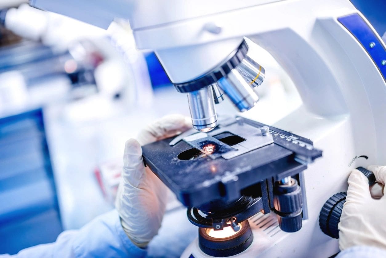
Advanced Imaging for Drug Development
Collaborating to Advance Structural Insights for Therapeutics
Introduction
A mid-sized biotechnology company approached GOTBio to address a critical problem in the development of their novel cell therapy. The therapy centered around the manipulation of G-Protein-Coupled Receptors (GPCRs), which are key regulators of cellular communication and play a significant role in signal transduction across cell membranes. Specifically, the company was interested in understanding the structure and dynamics of a particular GPCR that formed a multicomponent adhesion contact with the extracellular matrix (ECM).
To solve this problem, GOTBio leveraged their extensive network and connected the company with a laboratory at the University of California (UC), which specialized in advanced imaging techniques. The lab utilized Total Internal Reflection Fluorescence (TIRF) microscopy to help the company visualize and quantify the movement and interaction of the GPCR at the cell membrane-ECM interface.
Objectives
- GPCR Structural Determination: Provide high-resolution imaging of the GPCR to understand its structural dynamics and confirm its role in the adhesion complex.
- Quantification of GPCR Movement: Use advanced microscopy to visualize the kinetics of GPCR movement within the cell membrane.
- Enhance Cell Therapy Development: Help the company develop their cell therapy by providing insights into how GPCRs function in the cellular environment, ultimately optimizing therapeutic design.
Approach
- Problem Scoping:
- GOTBio’s initial discussions with the biotech company revealed that the understanding of GPCR-ECM interactions was essential for advancing their therapeutic project. This was critical for designing cells that could interact with the ECM in predictable and beneficial ways.
- GOTBio identified a UC lab that had cutting-edge expertise in single-molecule imaging and TIRF microscopy, a technique that would allow the company to observe GPCR interactions in real time.
- Technology Selection:
- TIRF Microscopy: The UC lab suggested using TIRF microscopy, which provides a high-resolution view of molecular events occurring near the cell membrane. This method is ideal for studying receptor interactions at the interface between the membrane and ECM.
- The technique allowed researchers to illuminate only the region immediately adjacent to the membrane, reducing background noise and enhancing the clarity of the images.
- Experimentation:
- Sample Preparation: The UC lab collaborated with the company to prepare cells expressing the GPCR of interest. Cells were labeled with fluorescent markers to visualize the GPCR and the ECM components involved in the adhesion complex.
- Imaging Protocol: Using TIRF microscopy, the team captured images and videos of the GPCR in real-time as it interacted with ECM molecules. They collected data on GPCR clustering, diffusion rates, and conformational changes during adhesion events.
- Data Analysis: The lab used advanced image analysis tools to quantify the dynamics of GPCR movement, including tracking the speed and patterns of diffusion within the membrane. Kinetic models were generated to describe how the receptor responds to ECM cues.
- Results:
- The experiments provided clear images of GPCRs at the membrane-ECM interface, showing the formation of adhesion complexes in real-time.
- Kinetic Insights: The team successfully quantified the movement of GPCRs, demonstrating that the receptor exhibited distinct patterns of clustering and movement when in contact with the ECM. The data revealed the key regions of the receptor that were involved in adhesion, which provided valuable information for refining the cell therapy.
- Structural Data: High-resolution images allowed the biotech company to confirm the structural configuration of the GPCR within the adhesion complex, which was previously unknown. This helped them better understand the role of the receptor in their therapeutic strategy.
Impact and Conclusion
The collaboration between GOTBio, the UC lab, and the biotech company yielded significant insights into the structure and function of GPCRs at the membrane-ECM interface. By using TIRF microscopy, the UC lab provided the biotech company with critical data on GPCR movement and adhesion dynamics, which played a key role in refining their cell therapy development process.
The partnership exemplified how academic expertise in advanced microscopy can help solve real-world problems in the biotechnology industry, leading to more effective and targeted therapies. As a result, the company was able to make informed decisions on how to further develop their cell therapy, improving both the efficacy and safety of their product.
Key Takeaways
- Advanced Imaging for Drug Development: TIRF microscopy enabled the company to visualize GPCR interactions at the cellular level, a critical step in optimizing cell therapies.
- Collaborative Research: GOTBio acted as a facilitator, connecting the company with a specialized lab, fostering an academic-industry partnership that drove innovation.
- Quantitative Insights: The ability to quantify GPCR movement and interactions provided the company with valuable data that was pivotal for designing their therapy.
Future Directions
This collaboration opens the door for future projects involving the use of advanced imaging techniques to study other cell surface receptors and their roles in various therapeutic contexts. The success of this case suggests the potential for broader applications of TIRF microscopy in the biotechnology sector, especially for therapies involving receptor-mediated cell signaling.
Recent Posts
- The Importance of Marketing and Market Research in Biotech 12/10/2024
- How AI and Bioinformatics Are Revolutionizing the Biotech Landscape 12/10/2024
- The Role of Collaborative Intelligence in Biotech Innovation 12/10/2024
- Advanced Imaging for Drug Development 11/21/2024
- Solving Relocation Issues 11/21/2024
Categories
- Blogs (3)
- Business Case Studies (5)
- News articles (3)
- Scienctific Case Studies (8)

